
There
are simply some things in life that I don't understand.
The
Bible says God wouldn't give us what we couldn't handle...
Everyone
knows that I'm currently in Remission for Acute Promylocytic Leukemia...
But
I didn't realistically think about other cancers... especially ones that could
affect infertility, for female issues...
I
got my Ultrasound on June 3rd, and the results were anything but normal...
We
know that my HSG in January determined my fertility was not the greatest, and
that I had about a 5% chance of pregnancy... due to the large amount of
scarring on my endometrium, and my partially closed Fallopian tube... the
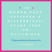
scarring was due to my large amount of miscarriages... I didn't really like
that doctor, but that is a different story... I plan to get a second opinion in
the future...
So
anyway... the ultrasound...
Gynecological
Report (Signed Final 06/03/2013 04:21 pm)
Patient
Info
------------
ID #:
091195
D.O.B.:
09/26/89 (23 yrs)(F)
Name:
APRIL DRIESSE
Visit
Date: 06/03/2013 10:47
Performed
By
------------
Performed
By:
Olesya
S Jungkman RDMS
Referred
By: GUDDETI
PALLAVI
MD
Location:
Manchester
Service(s) Provided
-------------------
GYN
PELVIC TRANSVAGINAL ONLY 76830
Indications
-----------
Polycystic
Ovarian Disease
History
-------
Age:
23
LMP:
05/19/13
Day
Of Cycle: 16
Menses:
Regular
Uterus
------
Uterus:
Present
Position:
Anteverted
Size
(cm) L: 7.88 W: 4.24 H: 3.38 Vol (ml): 59.1
Description:
Normal appearance
Endometrium
-----------
Endometrium:
Normal appearance
Thickness(mm):
5.62
Cervix
------
Normal
appearance
Cul-De-Sac
----------
No
fluid was Visualized
Right
Ovary
-----------
Status:
Visualized
Size
(cm) L: 3.31 W: 2.33 H: 1.96
Vol
(ml): 7.9
Morphology:
Polycystic
appearance
Left
Ovary
----------
Status:
Visualized
Size
(cm) L: 2.53 W: 2.44 H: 1.85 Vol (ml): 6
Morphology:
Polycystic appearance
Comment:
Echogenic area seen within the overy - 10 x 10 x 6mm.
Impression
----------
Anteverted
uterus 7.9 X 3.4 X 4.2 cm
Normal
myometrium
Endometrial
echo measures 5.6 mm in maximal dimension
Right
ovary measures 3.3 X 2 X 2.3 cm
Left
ovary measures 2.4 X 2.5 X 1.9 cm
central
small calcification
10mm X 6 mm unchanged in appearance from prior exam
6/12
Multiple
small peripheral ovarian follicles noted within both ovaries
No
free pelvic fluid
This ultrasound
report with graphs is available for review in our Faculty copy of AS Software Inc. Colleen M Barber,
MD Electronically Signed Final Report 06/03/2013 16:21
Component Results:
There is no component information for this result.
General Information
Collected:
6/3/2013
12:31 PM
Resulted:
6/3/2013 4:22 PM
Ordered
By: PALLAVI GUDDETI, MD
Result
Status: Final result
These
results were automatically released. Please contact your provider’s office if
you have questions.
What
is concerning to me is this:
Calcification
in soft tissues occur when there is deposition of calcium salts in dead or
degenerated tissues. Tissues can degenerate in response to infection and
inflammation, in response to tumors, or in response to decreased blood flow to
that particular area. Calcification in ovaries is taken to be a sign of a
previous or present problem.
Previous
issues like infections can show up as calcification. Present issues like tumors,
benign or malignant, can also show up as calcification's.
For
many years, calcification in ovaries was believed to be due to cancers causing
degeneration of tissues. But presently, research has shown that benign lesions
are more common than malignant tumor.
Whatever
the cause, the standard treatment of a calcification in an ovary is surgery to
remove the ovary and submitting the ovary for pathological examination so that
a malignant lesion may not be missed.
An
MRI can also identify cancers to some extent since blood flow increases in a
cancerous tumor. A PET scan is even more diagnostic.
So NOW WHAT??
My labs in May showed my white blood cell increasing, my platelets and red blood cells decreasing, which was concerning, because these are signs that my body may not be in remission anymore.
These new ultrasound results have me wondering:
How can this calcification remain for a year?
Is it the reason why I can't get pregnant and when I do, am not able to sustain pregnancy?
I wonder these things, because my doctors think MTHFR and my clotting disorders are not affecting my fertility...
So I put a call into my OBGYN and their going to be ordering an endometrial byposy, to rule out Endometrial Cancer... because the above results signify that I could have Endometrial Cancer...
GREATTTTTTT.....
Just another thing that I need to add to my medical history at 24...
Just another thing that might cost me my female fertility, and take up that last 5% of chance that I may be able to be a mother to my fiancee's children... (Which by the way, we were looking forward to, he said to me last night "I was actually looking forward to you being the mother to my next child(ren) and now this is discerning but hey, if that's the case, we have 3 beautiful kids we can take care of (he has 2 from a prior relationship, and then Gabby) and while I understand where he is coming from... I'm quite disheartened because I am wanting to be a mother to his children... I intend to marry him after-all .. and plan to fight this battle... and pray that God bless me with another child...


So now we wait...

 Born: June 25th 2009


1 comments:
:( i really hope that its not cancer
Post a Comment